Dental and Maxillofacial Radiology.
During specialty training in dental and maxillofacial radiology, trainees develop skills in reporting dental plain radiographs and Cone Beam CT, as well as performing diagnostic and interventional ultrasound and sialography. Additionally, we have relationships with radiology units throughout the region to allow trainees to gain experience in head and neck cross-sectional imaging and contribute to multi-disciplinary meetings.
Training Programme Directors
Dr Edward Walker (All)
Further information on recruitment, ARCP, study leave and period of grace can be found here.
Training follows the Dental and Maxillofacial Radiology Specialty Training curriculum. The Diploma in Dental and Maxillofacial Radiology (DDMFR) examination is a requirement for satisfactory completion of specialist training. Entry to the DDMFR requires successful completion of the physics module of the First FRCR Examination in Clinical Radiology, which can be attempted from 6 months into training, and the completion of specific periods of UK specialist training in Dental and Maxillofacial Radiology. Preparation for the First FRCR examination is supported by attendance at the Leeds Radiology Academy, where trainees will undertake a formal lecture programme alongside the ST1 Clinical Radiology cohort.
The DDMFR exam consists of two parts; part A can be attempted from 12 months into training, and part B from 30 months into training.
All training consultants have honorary senior lecturer positions, and contribute to a number of different undergraduate and postgraduate university modules. Trainees will also be encouraged to contribute to these programmes and will have the opportunity to hold honorary clinical lecturer positions.
The department has several active research and innovation streams, predominantly working in collaboration with the Leeds Teaching Hospitals Oral and Maxillofacial Surgery department.
Edward Walker
Ed is the Training Programme Director for DMFR in the region. He completed DMFR training in 2021 and has been a consultant in the region since then. He works in both Leeds Teaching Hospitals and Mid Yorkshire Teaching Hospitals Trusts and has clinical interests across a broad range of imaging modalities, and contributes to the regional head and neck oncology multi-disciplinary team. Ed has a number of collaborative research interests with other specialties, especially around optimising radiology in patient specific implant planning pathways
Niall O'Neill
Niall is a consultant in dental and maxillofacial radiology based at the Leeds Dental Institute. He studied dentistry at Queen's University Belfast and undertook his specialty training at Guy's Hospital, London. Niall has a particular interest in minimally invasive management of obstructive salivary gland disease.
Brenda Murray
Brenda is a Consultant in Dental and Maxillofacial Radiology, Honorary Senior Lecturer and Clinical Director of Leeds Dental Institute. She qualified from Queen’s University in Belfast in 1998 after intercalating both a BSc (Hons) and PhD in Anatomy during her undergraduate training. Brenda then went onto complete a one year Research Fellowship in Belfast and was subsequently appointed as a Clinical Lecturer in Oral Sciences at the Queen’s University, Belfast. During this time she completed a Postgraduate Certificate in Higher Education Teaching. She came to Leeds as a Specialist Registrar in 2003 and was appointed as a consultant in Leeds Dental Institute in 2008. Brenda has held a number of educational roles. She has been Chair of the Specialist Advisory Committee for the Additional Dental Specialties and led the development of the new curriculum for speciality training in Dental & Maxillofacial Radiology. She has also been Training Programme Director and National Recruitment Lead for the speciality and a member of the speciality exam board of the Royal College of Radiologists. Her main clinical interests are in ultrasound of the head and neck and cone beam CT.
Rosalyn Clarkson
Rosalyn is a Consultant in Dental and Maxillofacial Radiology.
Dental and Maxillofacial Radiology
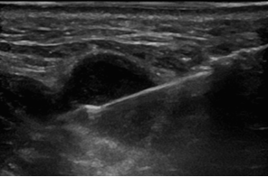
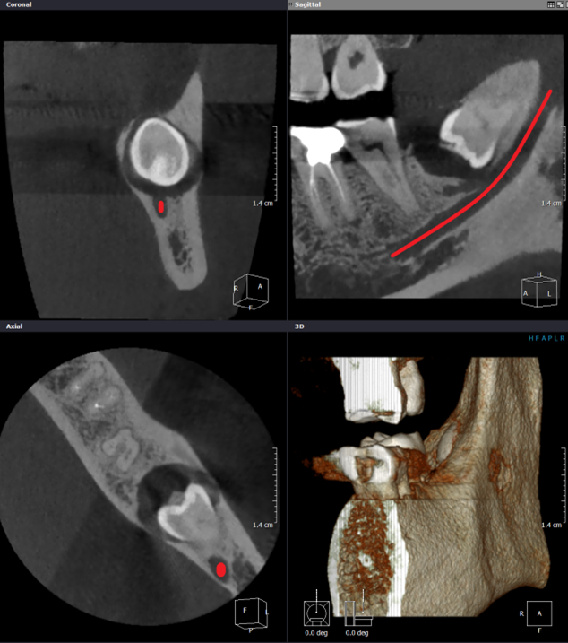
Dental and Maxillofacial Radiology is a rewarding and challenging career due to the diversity and complexity of the work. Yorkshire and the Humber offers fully integrated training in the both head & neck and dental components of the curriculum. Practically, this means that trainees are exposed to advanced imaging modalities and interventional procedures such as ultrasound guided interventions (figure 1), dental Cone Beam CT (figure 2) and MR imaging throughout training, ensuring that experience is gained in the full spectrum of the specialty. There are opportunities for training with both other dental speciality trainees and clinical radiology trainees and trainees have the benefit of access to the Leeds Radiology Academy.
Training is primarily based in Leeds Teaching Hospitals NHS Trust but will include placements in a number of other trusts in the region. This allows trainees to receive broad based training with involvement in local multi-disciplinary team and clinico-radiological meetings. Through the integration of placements at peripheral trusts, trainees’ work is supervised by, and feedback received from, a range of Consultant Dental & Maxillofacial Radiologists and Clinical Radiologists with a Head and Neck subspecialty interest.
A number of features of the training program have stood out to me:
- Active participation in regional Head and Neck oncology and Salivary Gland multi-disciplinary team meetings
- Day-to-day integration of Head & Neck and Dental components of the specialty from early in training
- Direct involvement in quality improvement and research projects
- Training with renowned specialists across multiple hospital trusts and sites
- Friendly, approachable supervisors and patient orientated teams at all trusts involved in the training program
Designed by E Walker, Dental and Maxillofacial Radiology Trainee 2021